Van Gieson's Picrofuchsin Staining: A Useful Tool in Histology
Van Gieson's picrofuchsin staining is a histological technique used to visualize the structure and composition of tissues, particularly collagen. This staining method has been widely used in biomedical research and diagnostics to study the morphology of various tissues, including skin, bone, and blood vessels. By highlighting the differences in tissue composition, van Gieson's picrofuchsin staining provides valuable information for researchers and clinicians to understand tissue function and disease mechanisms.
Understanding the Staining Process
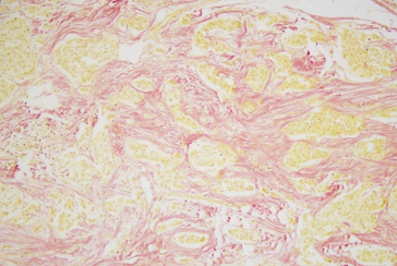
The van Gieson's picrofuchsin staining process involves treating tissue sections with a combination of picrofuchsin and van Gieson's stain. Picrofuchsin is a dye that stains collagen and other fibrillar proteins, while van Gieson's stain is a mixture of picric acid and acid fuchsin that stains other tissue components, such as cells and nuclei. The resulting stained tissue sections can be examined under a microscope to visualize the distribution and organization of collagen and other tissue components.

Applications in Research and Diagnostics
Van Gieson's picrofuchsin staining has a wide range of applications in biomedical research and diagnostics. One of the main uses of this staining method is to study the structure and composition of collagen-rich tissues, such as skin and bone. By examining the organization and distribution of collagen fibers, researchers can gain insights into tissue function and disease mechanisms, such as fibrosis and osteoporosis.
In addition to its use in basic research, van Gieson's picrofuchsin staining is also used in clinical diagnostics to examine tissue biopsies and diagnose various diseases, including cancer and inflammatory disorders. The staining method can help clinicians to visualize the extent of tissue damage and inflammation, and to identify specific cellular and tissue components that are involved in disease pathology.
Selecting the Right Staining Method
When selecting a staining method for histological analysis, researchers and clinicians need to consider several factors, including the type of tissue being examined, the specific research or diagnostic question being addressed, and the level of detail required. Van Gieson's picrofuchsin staining is a useful tool for visualizing collagen-rich tissues, but it may not be the best choice for examining other types of tissues or cellular components.

Implications and Future Directions
Van Gieson's picrofuchsin staining is a valuable tool in histology, providing valuable information about tissue structure and composition. As research and diagnostics continue to evolve, it is likely that new staining methods and techniques will be developed to visualize and analyze tissue components. However, van Gieson's picrofuchsin staining will likely remain a useful method for examining collagen-rich tissues and understanding tissue function and disease mechanisms.
In conclusion, van Gieson's picrofuchsin staining is a useful histological technique that provides valuable information about tissue structure and composition. Its applications in research and diagnostics are diverse, and it will likely continue to be an important tool in the field of histology.
Celnovte
Celnovte
Van Gieson’s Picro Fuchsin (VG) Staining. (a–h) VG Staining Of Four

Van Gieson’s picro fuchsin (VG) staining. (a–h) VG staining of four ...
Van Gieson's Stain | PDF

Van Gieson's Stain | PDF